Martial Arts
Jiu-Jitsu
Brazilian Jiu-Jitsu is a grappling art that heavily loads the grip, hips, and posterior chain. Guard work demands strong adductors and hip flexors, while passing requires explosive quadriceps and core. The forearms fatigue rapidly from gi grips, and the neck resists chokes and cranks under constant pressure.
Primary Muscles
Supporting Muscles
Primary Muscles
66Hand
Abductor digiti minimi of left hand
Located in the pinky side of the left palm, this muscle fans out the little finger for grip adjustments in sports or tools. It supports fine motor control and power grip stability. Climbers and grip athletes rely on it for pinky strength.
Hand
Abductor digiti minimi of right hand
In the right hand's hypothenar area, this muscle abducts the pinky finger, enhancing grip width for tools or sports. Crucial for musicians and weightlifters needing precise finger spread. Builds hand endurance.
Hand
abductor pollicis brevis
The thenar muscle at the thumb base abducts the thumb for pinch grips and opposition. Essential for fine tasks like texting or tools. Thumb trainers value it for dexterity.
Forearm
abductor pollicis longus
This forearm muscle runs to the thumb base, abducting and extending it for radial deviation. Vital for wrist stability in lifts like deadlifts. Forearm specialists train it for grip resilience.
Hand
adductor pollicis
Deep thumb adductor pulls thumb toward palm for key pinch strength. Vital for gripping keys or tools. Handstrength pros train it hard.
Forearm
brachioradialis
The brachioradialis is the thick forearm muscle on the radial (thumb) side, visible in hammer curls. It flexes the elbow in neutral grip, stabilizing during pulls and carries. Builds Popeye forearms for grip strength.
Forearm
extensor carpi radialis brevis
The ECRB is a forearm extensor on the radial side, extending and abducting the wrist. Mid-forearm location builds wrist stability for racquets and grips.
Forearm
extensor carpi radialis longus
The ECRL is the longer radial wrist extensor, more proximal in forearm for powerful extension and abduction. Stabilizes in heavy pulls.
Forearm
extensor carpi ulnaris
The ECU is the ulnar (pinky) wrist extensor in posterior forearm, balancing extension with deviation. Key for stability in sports.
Forearm
extensor digiti minimi
The EDM is a thin forearm muscle specifically extending the pinky finger at knuckles and wrist. Lies ulnar to EDM, aids fine grip control.
Forearm
extensor digitorum
The extensor digitorum is the central posterior forearm muscle, extending fingers 2-5 at knuckles and wrist. Fan-like tendons create dorsal hand ridges for grip extension.
Forearm
extensor indicis
The extensor indicis is a narrow forearm muscle that specifically extends the index finger (pointer finger), independent of the other fingers. It's essential for precise gripping and pointing motions in sports like tennis or climbing. Fitness pros value it for finger independence in grip training.
Forearm
extensor pollicis brevis
The extensor pollicis brevis sits in the forearm and extends the thumb at its base (metacarpophalangeal joint). It powers thumb opposition and pinch grips vital for weightlifting and climbing. For fitness, it's key in maintaining thumb stability during heavy pulls.
Forearm
extensor pollicis longus
Running deep in the forearm, the extensor pollicis longus straightens the thumb's end joint and adducts it. It's crucial for power grips in deadlifts and pinch strength. Athletes train it for thumb endurance in prolonged holds.
Forearm
flexor carpi radialis
Flexor carpi radialis is a forearm muscle that flexes and abducts the wrist toward the thumb side. Vital for hammering motions, pull-ups, and racket swings. Fitness staple for wrist strength in gymnastics and weights.
Hand
Flexor digiti minimi brevis of left hand
In the left hand's hypothenar eminence, this muscle flexes the pinky finger at its base for power grips. Key for musicians and rock climbers needing pinky strength. Enhances grip variety in fitness.
Hand
Flexor digiti minimi brevis of right hand
Right hand hypothenar muscle flexing pinky MCP joint for grip power. Essential for right-handed tools.
Lower Leg
flexor digitorum longus
The flexor digitorum longus is a powerful muscle in the deep posterior compartment of the lower leg, running from the tibia down to the toes. It flexes the four smaller toes and assists in plantarflexion at the ankle, helping with push-off during walking, running, and jumping. Strong flexors like this are crucial for balance, propulsion, and preventing foot drop in athletes.
Forearm
flexor digitorum profundus
Located in the deep anterior forearm, the flexor digitorum profundus flexes the distal joints of fingers 2-5, enabling a strong grip. It's vital for power activities like rock climbing, weightlifting, or crushing a grip trainer. Weakness here leads to dropped fingers and poor hand function.
Forearm
flexor digitorum superficialis
This superficial forearm muscle flexes the middle joints of fingers 2-5, key for precise hand control in sports like tennis or guitar playing. It sits in the anterior forearm, aiding in everything from typing to throwing. Balanced strength prevents imbalances with extensors.
Hand
flexor pollicis brevis
This thumb muscle in the thenar eminence flexes the thumb's base, critical for pinching and precision grips like turning keys or holding tools. It's key for hand strength in weightlifting or racket sports. Imbalances lead to thumb weakness.
Forearm
flexor pollicis longus
Deep in the anterior forearm, flexor pollicis longus flexes the thumb's tip joint, powering thumbs-up gestures and strong grips. Vital for tools, phones, and sports requiring thumb control. Essential for fine motor strength.
Hand
Flexor retinaculum of left wrist
The flexor retinaculum (transverse carpal ligament) is a thick band across the palmar wrist forming the carpal tunnel roof. It holds flexor tendons in place during wrist motion, vital for grip strength in fitness. Imbalances affect hand endurance.
Hand
Flexor retinaculum of right wrist
The right wrist's flexor retinaculum secures flexor tendons through the carpal tunnel, enabling smooth wrist flexion in daily and athletic tasks. It supports grip integrity during pulls and presses. Dysfunction leads to hand fatigue.
Forearm
Humeral head of left flexor carpi ulnaris
The humeral head of the left flexor carpi ulnaris is the upper arm-origin portion of this forearm muscle on your left side, running from the inner elbow down to your wrist. It flexes and adducts the wrist, stabilizing it during gripping and weight-bearing activities. Strong FCU matters for forearm endurance in sports like tennis or climbing.
Forearm
Humeral head of left pronator teres
The humeral head of the left pronator teres originates from your left inner elbow and crosses to the mid-forearm, rotating the forearm palm-down. It's key for turning motions in daily tasks and sports like golf or throwing. Building it prevents forearm fatigue and rotation imbalances.
Forearm
Humeral head of right flexor carpi ulnaris
The humeral head of the right flexor carpi ulnaris starts at the inner elbow on your right side and runs to the wrist, enabling wrist bend and pinky-side tilt. Vital for right-handed grip strength in tools, weights, or racquets. It supports unilateral forearm power in asymmetric training.
Forearm
Humeral head of right pronator teres
This right-side humeral head of pronator teres links inner right elbow to mid-forearm, powering palm-down rotation for throwing, swinging, or tool use. Essential for right-dominant athletes to balance forearm rotators and avoid overuse.
Foot
Opponens digiti minimi of left foot
Small intrinsic foot muscle opposing pinky toe for transverse arch stability. Aids push-off in sprinting. Rarely isolated but supports forefoot dexterity in climbers.
Hand
Opponens digiti minimi of left hand
Hypothenar muscle opposing pinky finger to cup the palm for grip strength. Enhances power grip in weightlifting and climbing. Weakness affects fine motor tasks.
Foot
Opponens digiti minimi of right foot
The opponens digiti minimi is a small intrinsic muscle in the sole of the right foot that helps oppose the little toe against the other toes. It plays a key role in fine motor control for foot dexterity, which is vital for balance during activities like running on uneven terrain or gripping with toes in climbing. Fitness enthusiasts appreciate it for enhancing foot stability and preventing injuries in high-impact sports.
Hand
Opponens digiti minimi of right hand
This small muscle in the hypothenar eminence of the right hand enables opposition of the little finger, crucial for powerful grips like holding tools or barbells. It's essential for hand dexterity in weightlifting and climbing. Strengthening it improves overall grip strength and prevents hand fatigue.
Hand
opponens pollicis
The opponens pollicis sits at the base of the thumb in the thenar eminence, rotating the thumb to oppose the fingers for pinching and gripping. Vital for weightlifters handling barbells or dumbbells securely. It enhances precision in fitness activities like kettlebell work.
Forearm
palmaris longus
The palmaris longus is a slender forearm muscle running from elbow to palm, aiding wrist flexion and tensing the palmar aponeurosis for grip. Absent in 14% of people, it's key for climbers and grip athletes. It contributes to forearm endurance in pulling exercises.
Forearm
pronator quadratus
Deep forearm muscle between radius/ulna pronating the forearm (palm down) for screwdriver grips. Key for wrist stability in deadlifts. Enhances rotational forearm strength.
Hand
Set of dorsal interossei of left hand
These four small fan-shaped muscles on the back of the left hand abduct the fingers away from the middle finger. They work with other intrinsics for fine motor control and grip strength. Important for grip-intensive fitness like climbing or weightlifting.
Hand
Set of dorsal interossei of right hand
These four small fan-shaped muscles on the back of the right hand abduct the fingers away from the middle finger. They enable precise finger spreading and grip control. Crucial for hand-intensive sports and weight training.
Hand
Set of lumbricals of left hand
Worm-like muscles in left hand palm flexing knuckles while extending fingers for writing/gripping. Essential for fine motor fitness tasks.
Hand
Set of lumbricals of right hand
Same as left but right hand; key for dexterity.
Hand
Set of palmar interossei of left hand
Three muscles in left palm adducting fingers toward middle. Grip power for fitness.
Hand
Set of palmar interossei of right hand
Right palm adductors for grip.
Forearm
supinator
The supinator wraps around the upper forearm, supinating (turning palm up) the radius over the ulna. Essential for twisting motions in sports and daily lifts. Key for forearm endurance in grip-heavy training.
Foot
Third lumbrical of left foot
The third lumbrical of the left foot is a small worm-like muscle flexing the proximal toes for balance. Specific to left foot 3rd-4th toe interspace. Aids push-off in running/squats.
Foot
Third lumbrical of right foot
The third lumbrical of the right foot flexes proximal and extends distal phalanges of 4th toe for propulsion. Right foot specific. Enhances foot stability in unilateral training.
Forearm
Ulnar head of left flexor carpi ulnaris
The ulnar head is the larger part of the left flexor carpi ulnaris, on the medial forearm flexing and adducting the wrist. Essential for grip strength, hammering, and racket sports. Balances forearm for injury prevention.
Forearm
Ulnar head of left pronator teres
The ulnar head forms the deep ulnar part of the left pronator teres, pronating the forearm (palm down). Key for twisting motions like turning doorknobs or tennis serves. Supports grip and elbow stability.
Forearm
Ulnar head of right flexor carpi ulnaris
The ulnar head is the main portion of the right flexor carpi ulnaris on inner forearm, flexing and ulnar deviating wrist. Vital for strong grips in pulls and sports.
Forearm
Ulnar head of right pronator teres
Ulnar head of right pronator teres deep in proximal forearm, drives pronation. Important for rotational power.
Hip
gemellus inferior
Small deep rotator in the hip, gemellus inferior laterally rotates and stabilizes the thigh. Works with piriformis in squats and pivots. Crucial for hip control in sports.
Hip
gemellus superior
Tiny hip muscle above ischial spine, gemellus superior laterally rotates femur. Assists in twisting sports and hip stability. Part of deep six rotators.
Hip
gluteus maximus
Largest hip muscle, gluteus maximus extends and externally rotates thigh for powerful hip thrust in squats, deadlifts, running. King of posterior chain.
Hip
gluteus medius
Side hip muscle for abduction and stabilization during single-leg stance in running, squats. Prevents Trendelenburg gait.
Hip
gluteus minimus
Deepest glute under medius, minimus abducts and internally rotates hip. Key stabilizer for balance in yoga, hiking.
Hip
piriformis
The piriformis is a deep hip rotator in the glutes, externally rotating the hip for balance in squats and deadlifts. Tightness often causes sciatica-like pain. Essential for hip mobility in athletes.
Hip
quadratus femoris
Deep hip external rotator between ischium/femur for leg stability in single-leg work. Prevents twisting injuries in sports.
Thigh
biceps femoris
The biceps femoris is the lateral hamstring on the back of the thigh, with long and short heads forming a thick band from hip to knee. It flexes the knee, extends the hip, and rotates the leg outward, vital for running, jumping, and deadlifts. Key for posterior chain power and injury prevention.
Thigh
gracilis
Long, thin medial thigh muscle adducting and flexing knee. Aids cutting movements in soccer, stabilizing in squats.
Lower Leg
popliteus
The popliteus is a small knee muscle behind the joint that 'unlocks' the knee from full extension for flexion. Crucial for downhill running and pivoting in sports. Prevents knee hyperextension.
Thigh
semimembranosus
The semimembranosus is a posterior thigh hamstring forming the teardrop at knee back, flexing knee and extending hip. Powers deadlifts, lunges, and deceleration in sports. Prevents ACL strains by stabilizing.
Thigh
semitendinosus
Semitendinosus is the slender medial hamstring with a long tendon, flexing knee and extending hip. Aids in medial knee stability for cutting sports. Complements semimembranosus for balanced posterior chain.
Abdomen
Diaphragm
The diaphragm is the dome-shaped breathing muscle separating chest from abdomen, contracting to inhale. Central tendon anchors it, essential for core bracing in lifts and endurance.
Pelvis
External anal sphincter
The external anal sphincter is a skeletal muscle ring around the anus that you can consciously control for bowel movements. It maintains continence during daily activities and is strengthened via Kegels for pelvic floor health. Important for athletes in high-impact sports to prevent incontinence.
Abdomen
external oblique
External obliques form the outer 'V' of your abs on each side, rotating and side-bending the torso while compressing the abdomen. They're powerhouse muscles for rotational power in sports like golf, boxing, and throws. Train them for a defined waist and core stability.
Hip
iliacus
The iliacus is the fan-shaped hip flexor filling your pelvis bowl, partnering with psoas to lift the thigh. Crucial for running, kicking, and rising from sits. Tight iliacus contributes to anterior pelvic tilt and lower back strain in fitness enthusiasts.
Abdomen
psoas major
The psoas major is a deep hip flexor from spine to thigh, lifting knees in running/squats. Core to posture and power; tightness causes low back pain.
Pelvis
pubococcygeus
Part of pelvic floor, the pubococcygeus supports bladder/bowel, aids continence and core stability. Crucial for intra-abdominal pressure in heavy lifts like squats.
Supporting Muscles
101Hip
adductor brevis
Short inner thigh muscle adducts the thigh and assists rotation, key for lateral stability in squats and sprints. Middle adductor, it prevents groin pulls in dynamic sports. Essential for balanced leg power.
Hip
adductor longus
Prominent inner thigh muscle adducts and flexes the hip, powering lateral lunges and cutting moves. Prone to strains but key for athletic power. Gym staple for groin strength.
Hip
adductor magnus
Largest inner thigh muscle, with adductor and hamstring-like parts, adducts and extends hip powerfully. Mimics hamstring in deadlifts. Crucial for posterior chain balance.
Hip
adductor minimus
Small deep inner thigh muscle above magnus, aids adduction and rotation. Stabilizes hip in single-leg work. Often overlooked but prevents imbalances.
Thigh
gracilis
Long, thin medial thigh muscle adducting and flexing knee. Aids cutting movements in soccer, stabilizing in squats.
Hip
obturator externus
Deep hip external rotator and adductor originating from the pelvis obturator foramen. Stabilizes hip in deep squats and lateral movements. Often overlooked but prevents groin strains in agility training.
Hip
pectineus
The pectineus is a flat muscle on the inner upper thigh that flexes and adducts the hip, helping drive knees up in running or squats. It's crucial for lower body power in athletes. Tightness contributes to groin strain prevention.
Thigh
sartorius
The sartorius is the longest muscle in the body, spiraling from hip to inner knee, forming a 'tailor's muscle' for crossing legs. It flexes, abducts, and rotates the hip plus flexes the knee, key for soccer kicks and agility drills. Balances quad-dominant training.
Thigh
rectus femoris
The rectus femoris is the central quad muscle crossing both hip and knee, visible as the teardrop above your knee. It flexes the hip and extends the knee, powering sprints, jumps, and squats. Balanced development prevents knee pain in athletes.
Thigh
vastus intermedius
Deep central quad muscle under rectus femoris, extends knee powerfully. Core quad for squats and jumps.
Thigh
vastus lateralis
Largest quad on outer thigh, massive knee extender for lateral stability in lunges and sprints.
Thigh
vastus medialis
Teardrop inner quad (VMO) stabilizes patella, key for knee health in deep squats.
Upper Arm
coracobrachialis
The coracobrachialis is a small shoulder flexor deep in the upper arm, bridging coracoid to humerus. It flexes and adducts the arm, stabilizing shoulder in presses. Adds inner arm density for balanced delts.
Shoulder
deltoid
The deltoid is the rounded shoulder cap muscle with anterior, middle, and posterior fibers covering the shoulder joint. It abducts, flexes, and extends the arm for raises and presses. Builds the V-taper and protects the rotator cuff.
Shoulder
infraspinatus muscle
Infraspinatus caps the back of the shoulder blade, externally rotating the arm for throwing and serving. Key rotator cuff muscle for shoulder stability in presses and pulls.
Chest
serratus anterior
Serratus anterior wraps from ribs to scapula, protracting and upwardly rotating the shoulder blade for punches and presses. 'Boxer's muscle' prevents winging, crucial for overhead athletes.
Shoulder
subscapularis
The subscapularis is the anterior rotator cuff muscle filling the scapula's subscapular fossa. It internally rotates the arm and stabilizes the shoulder joint. Crucial for fitness in presses, pulls, and preventing dislocations.
Shoulder
supraspinatus
The supraspinatus tops the rotator cuff on scapula's fossa, initiating shoulder abduction. Vital for overhead presses and impingement prevention in weight training.
Shoulder
teres major
The teres major is a thick muscle from lower scapula to humerus, adducting and internally rotating the arm. 'Lat's little helper' for pulling exercises like rows and pull-ups.
Shoulder
teres minor
The teres minor is a narrow rotator cuff muscle on scapula's lateral border, externally rotating and stabilizing the shoulder. Key for throwing and pressing without winging.
Upper Arm
biceps brachii
The biceps brachii is the iconic front-of-arm muscle with two heads, located on the front of the upper arm from shoulder to elbow. It flexes the elbow and supinates the forearm, powering curls and underhand pulls. Essential for arm strength and aesthetics in any fitness routine.
Upper Arm
brachialis
The brachialis lies deep under the biceps in the lower front upper arm, the true powerhouse for elbow flexion. It works silently in every curl, providing thick arm mass without the peak. Crucial for heavy pulling and functional strength.
Neck
arytenoid cartilage
Paired laryngeal cartilages in throat enabling voice production via vocal cord movement. Not muscle but closest for neck; singers train supporting muscles. Vital for breathing/speaking.
Neck
cervical rotator
Cervical rotators are deep neck muscles like obliquus capitis inferior and splenius cervicis that turn the head side-to-side. Located deep in the upper cervical spine, they enable rotation for looking over shoulder. Vital for neck mobility in sports and daily turns.
Neck
cricothyroid
The cricothyroid is a small intrinsic laryngeal muscle in the anterior neck, tensing vocal cords for higher pitch. Located between cricoid and thyroid cartilages, it's key for singing and speaking. Matters for vocal athletes like singers.
Neck
digastric
The digastric has anterior and posterior bellies under the jaw, opening the mouth by depressing mandible. Runs from mandible to mastoid via sling, key for chewing and yawning.
Head
genioglossus
Fan-shaped tongue muscle from chin to tongue base, genioglossus protrudes and depresses tongue. Vital for swallowing, speech, and breathing in fitness vocal training.
Neck
geniohyoid
Thin neck muscle under chin pulling hyoid forward/up, geniohyoid aids swallowing and tongue movement. Supports neck stability in planks.
Neck
hyoglossus
The hyoglossus is a thin tongue muscle running from the hyoid bone (under chin) up into the tongue's side. It depresses and retracts the tongue, aiding swallowing and speech. Rarely targeted in fitness, but dysfunction affects eating and breathing mechanics.
Neck
iliocostalis cervicis
Iliocostalis cervicis is the neck portion of the erector spinae, running vertically along upper back to cervical ribs. It extends and laterally bends the neck, vital for posture in overhead lifts and sports. Supports head stability in fitness.
Head
inferior oblique
Inferior oblique is an eye muscle under the eyeball, rotating it up and out. Matters for gaze stability in dynamic sports; strains rare but affect tracking.
Neck
inferior pharyngeal constrictor
Inferior pharyngeal constrictor wraps lower throat, constricting for swallowing. Supports airway protection.
Neck
lateral crico-arytenoid
Lateral crico-arytenoid closes vocal folds for voice and swallow.
Neck
lateral thyrohyoid ligament
Lateral thyrohyoid ligament connects thyroid to hyoid, stabilizing hyoid in swallow. Not a muscle; passive.
Head
levator palpebrae superioris
The levator palpebrae superioris is a small muscle located above the eye within the orbit that elevates the upper eyelid. It plays a crucial role in opening the eyes for clear vision during workouts and daily activities. Fitness enthusiasts care about it because eyelid fatigue or weakness can impair focus during intense training sessions.
Neck
levator scapulae
The levator scapulae runs from the upper neck vertebrae to the top of the shoulder blade, lifting the scapula toward the head. It's key for shrugging motions and stabilizing the shoulder during overhead lifts. Gym-goers target it indirectly through shrugs and neck training to prevent shoulder hikes and imbalances.
Head
levator veli palatini
This small muscle in the side of the throat lifts the soft palate during swallowing and speech. It helps seal off the nasal cavity for proper voice resonance and prevents food from entering the nose. Relevant for singers or those doing breathwork in fitness to maintain clear airways.
Neck
longissimus capitis
Part of the erector spinae group, the longissimus capitis extends from the upper back along the neck to the skull, turning and tilting the head. It stabilizes the head during heavy lifts like deadlifts. Strong neck extensors prevent whiplash and support posture in athletes.
Neck
longissimus cervicis
This erector spinae muscle spans the neck vertebrae, extending and stabilizing the cervical spine. It keeps the head aligned during upright posture and dynamic movements like cleans. Vital for neck resilience in contact sports and heavy training.
Neck
longus capitis
A deep anterior neck flexor, the longus capitis flexes the head forward and stabilizes cervical vertebrae. It counters posterior muscles during neck crunches or wrestling bridges. Key for balanced neck strength and preventing forward head posture.
Neck
longus colli
The longus colli is the primary deep neck flexor, spanning all cervical vertebrae to flex and stabilize the neck. Crucial for chin tucks and preventing slouched posture in long training sessions. Builds neck endurance for grapplers and cyclists.
Neck
Median thyrohyoid ligament
Not a muscle but a ligament connecting thyroid and hyoid bones in the midline neck, it supports hyoid elevation during swallowing. Indirectly aids strap muscles in fitness for better airway control. Rarely targeted but important for throat stability.
Neck
middle pharyngeal constrictor
This throat muscle constricts the pharynx during swallowing, forming the middle band around the airway. Supports powerful swallows in athletes consuming large meals or shakes. Maintains throat tone for vocal endurance.
Neck
mylohyoid
The mylohyoid forms the floor of the mouth, elevating the hyoid and floor during swallowing and speech. It supports tongue movement for chewing tough foods in bulking diets. Key for jaw stability in powerlifters.
Neck
oblique arytenoid
Paired muscles on the back of the larynx that close the vocal folds during phonation and protect airways. Aids forceful coughs and Valsalva in heavy lifts. Important for vocal control in coaches yelling sets.
Neck
obliquus capitis inferior
Deep suboccipital muscle rotating the atlas on axis for head turns. Stabilizes craniocervical junction during neck twists in sports. Critical for proprioception and dizziness prevention in rotational training.
Neck
obliquus capitis superior
Upper suboccipital extending and bending the head laterally while stabilizing occiput. Works with rotators for precise head control in aiming sports. Prevents atlanto-occipital instability.
Neck
omohyoid
Strap-like muscle with two bellies depressing the hyoid during swallowing and speech. Tautens neck skin and aids large bolus swallows in athletes. Prevents hyoid elevation overload.
Neck
platysma
The platysma is a thin sheet-like muscle under the chin and neck skin, depressing the jaw and lower lip for expressions. Helps in neck stability during shrugs. Fitness note: resists 'neck bands' with age.
Neck
posterior crico-arytenoid
The posterior crico-arytenoid is the sole abductor of the vocal folds, essential for breathing by opening the airway. Vital for singers and athletes needing vocal control. Weakness affects breathing endurance.
Neck
rectus capitis anterior
Short neck flexor at base of skull flexing upper cervical spine for head nod. Stabilizes head in shrugs, prevents forward head posture.
Neck
rectus capitis lateralis
Lateral upper cervical flexor stabilizing side-bending head for gaze control. Aids neck endurance in upright posture.
Neck
rectus capitis posterior major
The rectus capitis posterior major is a small deep neck muscle at the base of your skull, running from the upper cervical spine to the occiput. It extends and rotates the head, helping maintain upright posture during daily activities and workouts. Strong suboccipitals like this one prevent forward head posture common in desk workers and lifters.
Neck
rectus capitis posterior minor
This tiny deep neck muscle sits just below the occiput, connecting the atlas to the skull. It assists in head extension and rotation, crucial for stabilizing your head during heavy lifts or prolonged sitting. Weakness contributes to chronic neck stiffness in fitness enthusiasts.
Neck
scalenus anterior
The anterior scalene anchors from neck vertebrae to first rib, aiding neck flexion and arm elevation. It lifts the first rib during deep breaths, important for overhead athletes. Tension contributes to thoracic outlet issues.
Neck
scalenus medius
The middle scalene runs from cervical transverse processes to first rib, assisting neck lateral flexion and rib elevation. Crucial for breathing during intense workouts and shoulder stability. Often tight in cyclists and throwers.
Neck
scalenus posterior
The posterior scalene attaches lower cervical vertebrae to second rib, flexing neck and aiding inhalation. Supports posture in endurance sports by stabilizing ribs. Less commonly injured but tightens with chronic coughs.
Neck
semispinalis capitis
This thick muscle spans upper thoracic and lower cervical to occiput, extending and rotating the head. Buffers heavy axial loads in powerlifters and stabilizes during shrugs. Key for countering forward head in tech users.
Neck
semispinalis cervicis
The semispinalis cervicis runs from upper thoracic to cervical transverse processes, extending the neck. Supports head during upright posture and overhead presses. Vital for cervical stability in contact sports.
Neck
Set of anterior cervical intertransversarii
These small muscles run between the transverse processes of adjacent cervical vertebrae in the front of the neck. They assist in lateral flexion and slight rotation of the neck, helping maintain proper head alignment during movement. Fitness enthusiasts care because they support neck stability in exercises like overhead presses or deadlifts.
Neck
Set of interspinales cervicis
Tiny paired muscles between spinous processes of cervical vertebrae, aiding neck extension and stabilization. They help maintain posture during upper body lifts. Relevant for neck strength in fitness routines.
Neck
Set of posterior cervical intertransversarii
Back neck muscles between transverse processes for lateral flexion/rotation opposite to anterior.
Neck
splenius capitis
Broad muscle upper neck/shoulder blade extends/rotates head. Key for neck strength in presses.
Neck
splenius cervicis
Lower splenius extends/rotates cervical spine.
Neck
sternocleidomastoid
Prominent neck muscle turns/tilts head, flexes neck, aids breathing. Visible in fitness for posture/aesthetics.
Neck
sternohyoid
The sternohyoid is a thin, strap-like muscle in the anterior neck that runs from the sternum to the hyoid bone. It depresses the hyoid bone during swallowing and speaking, stabilizing the larynx. For fitness enthusiasts, it's key for neck stability in exercises like deadlifts or overhead presses, preventing strain during heavy breathing.
Neck
sternothyroid
The sternothyroid lies deep in the anterior neck, connecting the sternum to the thyroid cartilage. It depresses the larynx for swallowing and voice modulation. Fitness folks value it for neck resilience during compound lifts or contact sports.
Neck
stylohyoid
The stylohyoid is a slender muscle from the skull's styloid process to the hyoid bone in the upper neck. It elevates and retracts the hyoid during swallowing. Important for fitness in maintaining neck posture during dynamic movements like cleans.
Neck
stylohyoid ligament
The stylohyoid ligament is a fibrous band connecting the skull's styloid process to the hyoid bone, providing passive stability in the upper neck. It doesn't contract but supports hyoid movement during swallowing. Relevant for fitness in assessing neck tension from poor posture.
Neck
stylopharyngeus
The stylopharyngeus elevates the pharynx and larynx from the skull's styloid to the pharyngeal wall. It aids swallowing and speech by lifting the throat. Fitness relevance: supports airway patency during intense workouts.
Head
superior oblique
The superior oblique is an extrinsic eye muscle originating near the nose, passing through a pulley to depress and intort the eyeball. It controls downward/outward gaze. Fitness note: eye stability aids visual tracking in sports.
Neck
superior pharyngeal constrictor
The superior pharyngeal constrictor forms the upper pharynx wall, constricting to propel food during swallowing. Originates from skull base to pharynx. Supports safe swallowing in athletes under exertion.
Head
Tendon of right levator palpebrae superioris
The tendon of the right levator palpebrae superioris attaches the muscle to the eyelid, elevating the upper lid for vision. Specific to right eye. Aids blink-free focus in precision sports.
Neck
thyro-arytenoid
The thyro-arytenoid adjusts vocal ligament tension inside larynx for pitch. Key for vocal athletes like singers.
Neck
thyrohyoid
Thyrohyoid elevates thyroid or depresses hyoid for swallow. Short neck muscle.
Neck
thyrohyoid membrane
Fibrous sheet between thyroid and hyoid, stabilizing larynx.
Neck
Transverse arytenoid
The transverse arytenoid is a single small muscle between the arytenoid cartilages in your larynx (voice box). It pulls them together to close the vocal folds for phonation and swallowing. Vital for voice production and airway protection, though not directly trained in fitness.
Head
Trochlea of left superior oblique
The trochlea is a cartilage pulley at the medial eye socket for the left superior oblique muscle tendon. It redirects the tendon to enable eye intorsion, depression, and abduction. Critical for coordinated eye movements, though not a muscle itself.
Head
Trochlea of right superior oblique
The trochlea is a cartilage pulley at the medial orbit for the right superior oblique muscle tendon. It enables precise eye intorsion, depression, and abduction. Key for binocular vision coordination.
Head
Uvular muscle
The uvular muscle elevates the uvula (soft palate dangler) during swallowing and speech. Aids gag reflex and velopharyngeal closure. Minor role in fitness but key for swallowing.
Neck
Vertical intermediate part of left longus colli
Vertical intermediate fibers of left longus colli flex neck laterally and forward. Deep neck stabilizer for posture.
Pelvis
iliococcygeus
The iliococcygeus forms the back part of the pelvic floor, spanning from pelvis to tailbone, supporting organs and pelvic stability. Important for core integrity in heavy lifts and preventing incontinence. Weakness shows in squats or postpartum.
Lower Back
iliocostalis lumborum
Iliocostalis lumborum is the lumbar erector spinae muscle along the back, from iliac crest to lower ribs, maintaining upright posture and spinal extension. Critical for deadlifts, squats, and anti-rotation core work.
Upper Back
longissimus thoracis
The longest erector spinae muscle, running parallel to the spine from sacrum to mid-back, it extends the spine and maintains upright posture. Essential for deadlifts, squats, and spinal stability in powerlifting. Weakness leads to back rounding under load.
Lower Back
lumbar rotator
Lumbar rotators (multifidus and rotatores lumborum) are deep spinal muscles that rotate and stabilize individual lumbar vertebrae. They prevent twisting injuries during rotational lifts like Russian twists. Essential for core stability in CrossFit and golf swings.
Upper Back
semispinalis thoracis
Semispinalis thoracis is a deep erector spinae muscle from thoracic spine, extending the vertebral column. Maintains thoracic posture during deadlifts and rows. Prevents slouching in prolonged sitting.
Lower Back
serratus posterior inferior
Deep muscle from lower thoracic spine to lower ribs, draws ribs down and back for exhalation. Aids breathing in heavy lifts like deadlifts.
Lower Back
Set of interspinales lumborum
Small muscles between lumbar spinous processes that extend and stabilize the lower back. Key for spinal integrity during squats and deadlifts. Fitness pros target them for back health.
Upper Back
Spinalis
Medial erector spinae column spanning thoracic/cervical, extends spine unilaterally/ bilaterally. Core for upright posture in lifts.
Pelvis
Tendinous arch of levator ani
The tendinous arch of levator ani is a fibrous white line on the pelvic sidewall anchoring levator ani muscles for pelvic floor support. It transmits fascia for stability during lifts. Critical for core integrity in squats and deadlifts.
Chest
Abdominal part of left pectoralis major
The abdominal part of the left pectoralis major is the lower portion of the left chest's primary pushing muscle, attaching from the sternum down to the ribs near the abdomen. It drives shoulder adduction and flexion, crucial for powerful presses like bench variations and dips. Fitness enthusiasts target it for that defined lower chest line in bodybuilding.
Chest
Abdominal part of right pectoralis major
The abdominal part of the right pectoralis major forms the lower section of the right chest's main power muscle, spanning from the sternum to the abdominal ribs. It excels in adduction and flexion for presses and flyes, key for balanced chest development. Gym-goers love it for carving out that teardrop lower chest aesthetic.
Chest
Anterior papillary muscle of right ventricle
This cardiac muscle inside the right ventricle anchors mitral valve leaflets for proper heart pumping. Not skeletal, but endurance training optimizes heart efficiency. Matters for cardio performance indirectly.
Chest
Anterolateral head of lateral papillary muscle of left ventricle
Specialized head of left heart papillary muscle securing mitral valve for left ventricle ejection. Critical for oxygenated blood flow. Athletes benefit from cardiac hypertrophy here.
Chest
External intercostal muscle
External intercostals are thin muscles between the ribs that elevate them during inhalation, aiding deep breathing for core stability in lifts. They're vital for endurance athletes needing efficient oxygen uptake. Fitness training enhances respiratory muscle power.
Chest
Innermost intercostal muscle
Innermost intercostals are deep rib muscles aiding forced expiration like coughing. Support breathing in high-intensity training.
Chest
Internal intercostal muscle
Internal intercostals between ribs assist expiration and stabilize chest in lifts.
Chest
Lateral papillary muscle of left ventricle
Papillary muscle in heart's left ventricle anchors mitral valve to prevent backflow. Endurance training strengthens indirectly.
Chest
pectoralis major
The massive pectoralis major covers the chest, powering arm adduction, flexion, and rotation for bench presses and push-ups. It's the prime mover for upper body pushing strength in fitness. Building it creates that powerful chest aesthetic.
Chest
pectoralis minor
Under the pec major, the pectoralis minor stabilizes the scapula, aiding shoulder protraction in dips and push-ups. Key for shoulder health in overhead athletes. It prevents winging scapula in fitness training.
Chest
transversus thoracis
The transversus thoracis are thin muscle bands on the inner chest wall beneath the sternum. They compress the chest during forced exhalation and stabilize the ribs. Important for deep breathing control in fitness and core stability.
Recommended Exercises
12

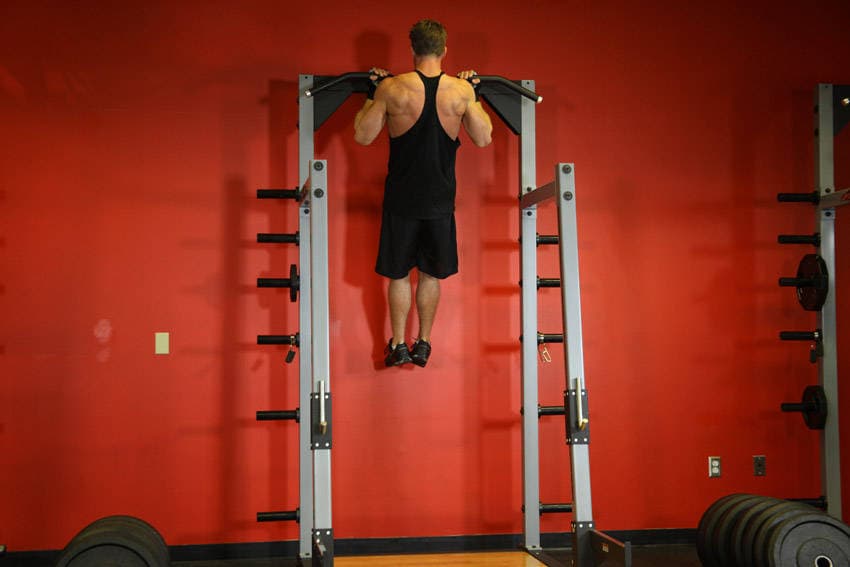
Pullups

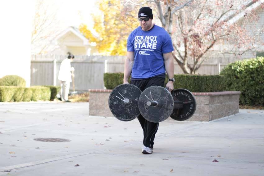
Farmer's Walk

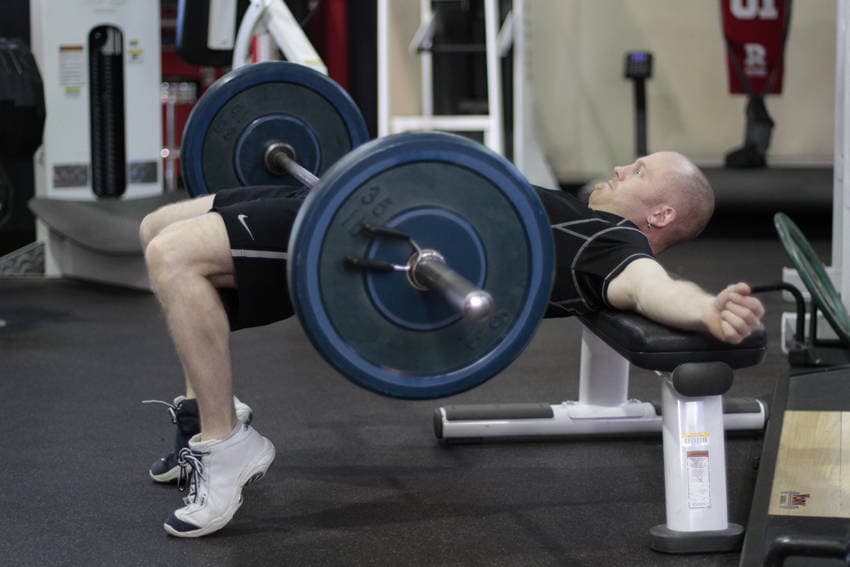
Barbell Hip Thrust

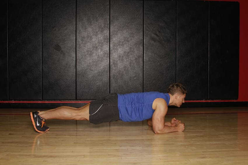
Plank

Chin-Up


3/4 Sit-Up


90/90 Hamstring


Ab Crunch Machine


Ab Roller


Advanced Kettlebell Windmill


Air Bike

